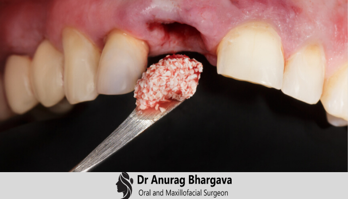
Bone graft is a small surgical procedure done in the dental room to build up new bone in the area of your jaw that is used to hold teeth. A small cut is made in your gum to expose the bone beneath it, and then grafting material is added. The grafting material is processed bone that serves as a platform, around which your body will deposit new bone cells. The grafting material will, in the end, be absorbed by your body and replaced by your new bone.
Bone Graft Process
The method for placing a bone graft requires anesthesia, though oral or IV sedatives can also be used to get relief. As a small cut in your gum tissue needs to be made to access the underlying bone that will receive the graft, you may undergo certain soreness in the area after the surgery, this can be treated by pain relievers as well as ice therapy after the procedure. Though you will soon be normal, it may take your body around seven months for bone maturation to take place to receive your dental implant.
The grafting material required can come from various sources. Sometimes it is from your own body. Quite often it is bone from an animal or human donor that is processed by a laboratory to make it sterile and safe. Grafting material can also be synthetic. It comes in a variety of forms: powder, granules, putty or a gel that can be injected through a syringe.
Over the years, the jawbone associated with missing teeth deteriorates. This leads to a condition in which there are quality and quantity of bone suitable for dental implants. In these situations, most patients are not suitable for the placement of dental implants. In such situations, orthognathic surgeons like Dr. Anuraga Bhargava perform a bone graft.
Nowadays we have the potential to grow the bone where needed. This not only gives us the chance to place implants of proper length and width, but it also allows us to restore functionality and aesthetic look.
Bone grafting can mend implant sites with improper bone structure due to previous extractions, gum disease or injuries. The bone is either taken from a tissue bank or your bone is taken from the jaw, hip or tibia.
Major bone grafts are performed to correct imperfections of the jaw. These imperfections may arise due to traumatic injuries, tumor surgery, or congenital defects. Big defects are rectified using the patient's bone. This bone is collected from several different sites depending on the size of the defect. The common donor sites include hip, skull and lateral knee. These procedures are routinely performed in a hospital. Oral and maxillofacial surgeons like Dr. Anurag Bharagava treats such defects.
Types of Bone Graft
The different types of bone graft are as follows:
1. Autogenous Bone Graft
Autogenous bone grafts also called autografts are made from your bone, taken from somewhere else in the body. The bone is gathered from the chin, jaw, lower leg bone, hip or the skull. Autogenous bone grafts are beneficial, in that the graft material is your live bone, meaning it contains living cellular elements that improve bone growth.
However, one drawback to the autograft is that it requires a second procedure to collect bone from elsewhere in the body. Depending on your condition, the second procedure may not be suggested.
2. Allogeneic Bone
Allogeneic bone or allograft is dead bone collected from a cadaver, then processed to extract the water via a vacuum. Like autogenous bone, allogeneic bone cannot produce new bone on its own. But it serves as a framework over which bone from the nearby bony walls can grow to fill the void.
3. Xenogenic bone
Xenogenic bone is extracted from the non-living bone of another species like a cow. The bone is processed at high temperatures to stop immune rejection and contamination. Xenogenic grafts serve as a framework for bone from the nearby area to grow and fill the void.
The xenogenic bone graft does not require a second procedure to collect your bone, as in the case of autografts. However, because of these options, bone regeneration may take longer and have a less predictable outcome.
Removal of teeth is essential because of pain, infection, bone loss, or due to a fracture in the tooth. The bone that holds the tooth in the socket is spoiled by disease or infection resulting in distortion of the jaw after the tooth is extracted. When teeth are extracted the nearby bone and gums can diminish and retreat very quickly, resulting in flaws and a subside of the lips and cheeks.
These jaw flaws can create major problems in performing restorative dentistry whether your treatment includes dental implants, bridges or dentures. Jaw malformation from tooth removal can be prevented and corrected by a procedure called socket preservation. Socket preservation can enhance your smile’s appearance and increase your chances for successful dental implants. Oral and maxillofacial surgeons like Dr. Anurag Bhargava perform such socket preservation.
Several procedures can be used to preserve the bone and reduce bone loss after an extraction. In one common procedure, the tooth is removed and the socket is filled with bone. It is covered with gum, an artificial membrane that allows your body's natural ability to correct the socket. With this technique, the socket heals, eliminating shrinkage and subside of the surrounding gum and facial tissues. The newly formed bone in the socket provides a base for an implant to replace the tooth. If your doctor has suggested tooth removal, be sure to ask if socket preservation is necessary. This is important if you are planning on replacing the front teeth.
4. Ridge Augmentation
Ridge augmentation is a dental procedure usually performed following tooth extraction. This procedure helps regenerate the natural contour of the gums and jaw that may have vanished due to bone loss from a tooth extraction.
The roots of teeth are surrounded by the alveolar ridge of the jaw. When a tooth is extracted an empty socket is left in the alveolar ridge bone. This empty socket will recover on its own, filling with bone and tissue. Many times when a tooth is removed, the bone surrounding the socket breaks and is not able to cure on its own. The previous dimensions of the socket will deteriorate.
Rebuilding the original height and width of the alveolar ridge may be required for dental implant placement or aesthetic purposes. Dental implants require bone to support their structure and a ridge augmentation can help restructure this bone to accommodate the implant.
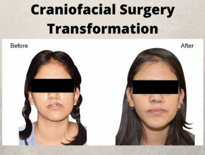
Bone Graft in Craniofacial Surgery
Bone defects in the craniomaxillofacial skeleton vary from the small periodontal defects to segmental defects resulting from trauma, surgical excision or cranioplasty. Such defects have complex three-dimensional structural needs, which are difficult to restore. In cranial vault flaws, the underlying brain needs permanent protection. Segmental jaw defects need restoration of mechanical integrity, temporomandibular joint function, and intermaxillary dental occlusion. Maintaining acceptable facial aesthetics is another important consideration in the treatment of facial defects, which cannot be underestimated. Bone grafts remain the perfect standard for reconstructing segmental bone defects.


